Post Doctorate
DR. GUETTER
- Doctor of Chiropractic, licensed in the State of Kentucky, License # 5291, 2011-Present
- Doctor of Chiropractic, Life University, Marietta, Georgia, 2011
- Internship, Apex Healthcare & Rehab, Lawrenceville, Georgia, 2009 – 2010
- National Board of Chiropractic Examiners, Part I, 2010
- National Board of Chiropractic Examiners, Part II, 2010
- National Board of Chiropractic Examiners, Part III, 2010
- National Board of Chiropractic Examiners, Part IV, 2011
- MRI Interpretation Review Qualified – Cleveland University, Kansas City
- Trauma Qualified – Cleveland University, Kansas City
- Expert Witness & Documentation Qualified – Cleveland University, Kansas City
- Hospital Qualified – Cleveland University, Kansas City
- Fellow, Primary Spine Care (Candidate)

MRI EXAMPLE
What is an MRI of the Spine?
Magnetic Resonance Imaging (MRI) is a noninvasive diagnostic imaging test doctors use to diagnose medical conditions. MRI uses a powerful magnetic field, radiofrequency pulses, and a computer to produce detailed pictures of internal body structures. MRI does not use radiation (x-rays). Detailed MR images allow doctors to examine the body and detect abnormalities and injuries. Currently, MRI is the most sensitive imaging test available for the spine.
MR imaging is used to assess or detect:
- Spine anatomy and alignment
- Birth defects in the vertebrae or spinal cord
- Trauma injury to the bone, disc, ligament, or spinal cord
- Disc and joint disease. Both are frequent causes of severe lower back pain and sciatica (back pain radiating into the lower leg)
- Compression or inflammation of the spinal cord and nerves
- Infection of the vertebrae, discs, spinal cord, or its coverings (meninges)
- Tumors in the vertebrae, spinal cord, nerves, or surrounding soft tissues
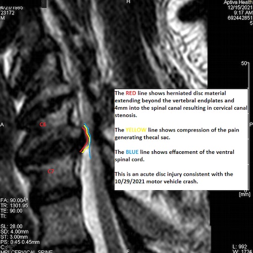
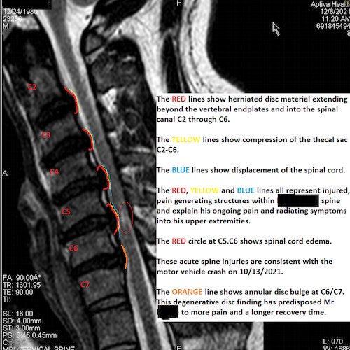
MRI IMAGE EXAMPLES